Cardiac Sarcoidosis and Myocarditis in Sarcoidosis: A Guide for Patients and Doctors
Introduction
Sarcoidosis is a chronic inflammatory disease of unknown cause that can affect any organ. The lungs, lymph nodes and skin are most often involved, but some people develop cardiac sarcoidosis, a potentially dangerous form in which inflammatory granulomas affect the heart muscle (myocardium). This can cause myocarditis, arrhythmias, heart failure and sudden death.
This complication often goes unrecognised, especially when symptoms are atypical. Early diagnosis requires awareness of specific and non-specific symptoms, careful interpretation of laboratory results and advanced imaging.
Typical and atypical symptoms
Typical symptoms include palpitations, chest pain, shortness of breath (especially on exertion), fatigue, dizziness or fainting, and peripheral oedema such as swollen ankles.
Atypical symptoms may include unexplained anxiety or inner discomfort, waking at night with breathlessness or needing to sleep semi-upright, non-specific back, shoulder or neck pain, variable blood pressure, mild headaches or blurred vision related to autonomic dysfunction, and pronounced fatigue after minor exertion. Because the symptoms vary so widely, many cases remain undiagnosed for a long time.
Biomarkers: what can blood tests tell us?
Troponin (hs-cTnT or hs-cTnI) is a highly sensitive indicator of heart-muscle injury. Even mildly elevated values may indicate active myocarditis, particularly in sarcoidosis, and can help monitor treatment response and relapse.
CK and CK-MB may provide additional information. CK-MB is more specific for cardiac muscle but less sensitive than troponin; total CK may also rise with skeletal-muscle inflammation.
ACE may indicate systemic sarcoidosis activity because granulomas produce it, but it is not specific. It is often normal in cardiac sarcoidosis and cannot be used to rule it out.
Serum calcium is elevated in about 10–20% of people with systemic sarcoidosis. It is less often elevated in cardiac disease, so a normal result does not exclude the diagnosis.
Diagnostic methods
An ECG records the heart’s electrical activity and may detect arrhythmias, atrioventricular block, pre-excitation or ventricular tachycardia.
Echocardiography assesses cardiac anatomy and function, including ejection fraction, chamber size and wall motion.
Cardiovascular magnetic resonance (CMR) is highly accurate for evaluating inflammation and fibrosis. Gadolinium contrast helps detect scars and oedema. MRI uses a magnetic field to image soft tissues without radiation.
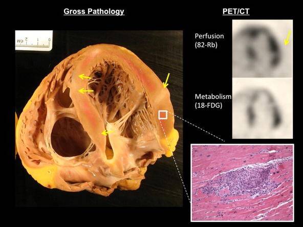
FDG-PET detects active inflammation using labelled glucose and can help assess treatment response and relapse.
Endomyocardial biopsy is rarely required. It can confirm granulomas, but its sensitivity is limited because the disease is focal and the procedure carries risks.
Monitoring people with suspected or confirmed cardiac sarcoidosis
Initial assessment may include blood tests (troponin, CK-MB, NT-proBNP, CRP, ACE and calcium), ECG and echocardiography, with CMR or PET to look for inflammation and fibrosis.
During treatment, troponin and CRP may be checked every 1–3 months, ECG every 3–6 months, and MRI or PET every 6–12 months or as clinically indicated. Symptoms should be monitored and immunosuppressive treatment adjusted when necessary; medicines may include corticosteroids, methotrexate or azathioprine.
Long-term follow-up includes regular cardiology visits, annual echocardiography, Holter ECG for suspected arrhythmias and, in people at high risk of sudden death, consideration of an implantable cardioverter-defibrillator (ICD).
Conclusion
In people with sarcoidosis, especially those with unexplained fatigue, palpitations or dizziness, cardiac sarcoidosis should be considered promptly. ACE and calcium may be elevated, but their value for detecting cardiac involvement is limited. High-sensitivity troponin and advanced methods such as CMR and PET provide more reliable information about inflammation and scarring. Diagnosis and treatment decisions must be made by the treating medical team.