

Schematic representation of the causes of sarcoidosis at the cellular level
How sarcoidosis develops
This text explains the connection between the development and outcome of sarcoidosis at the cellular level.
In simple terms—while those who are interested can read the detailed explanation:
1. A foreign body enters the body. We call this body an antigen. It most likely enters through the airways.
2. The immune system responds “too aggressively”. Small cells called macrophages are responsible for “eating” foreign material. Together they form an inflammatory cluster and create granulomas. During this defence process, macrophages may contribute to calcification of healthy tissue. For this reason, DO NOT TAKE CALCIUM OR VITAMIN D WITHOUT MEDICAL SUPERVISION, AND DO NOT TRY TO BOOST THE IMMUNE SYSTEM.
Lymphatic vessels, whose role is to remove toxins and reduce inflammation, become swollen as they encounter granulomas.
Treatment with glucocorticosteroids aims to reduce inflammation, while treatment with cytotoxic or immunosuppressive medicines such as methotrexate slows the immune response.
Detailed description:
Exposure to an unknown sarcoidosis antigen activates T cells and antigen-presenting cells (APCs), leading to the release of cytokines. CD4 activation can skew the immune system towards a Th1 or Th2 response, which in turn induces granuloma formation. At later stages, granuloma formation may be followed by resolution or by chronic disease with fibrosis.
A granuloma is a type of inflammation that occurs in response to different triggers, such as infections, foreign bodies or autoimmune disorders.
It is characterised by small clusters of cells that may develop in different parts of the body, including the lungs, skin and digestive system.
Granulomas consist of immune cells, such as macrophages, which accumulate in a particular area in response to a stimulus.
Sarcoid granuloma: It can develop in several organs, including the lungs, lymph nodes and skin. The cause of sarcoidosis is not fully understood, but it is believed to involve an abnormal immune response in which the immune system attacks the body’s own tissues. Sarcoid granulomas can cause symptoms such as cough, breathing difficulties and skin lesions.
Macrophages are a type of leukocyte found in humans and animals. They are microscopically larger than neutrophils and their main task is to clear waste from the body and fight foreign substances (antigens).
They develop from blood monocytes, which are produced in the bone marrow. Macrophages contribute to anthracosis and asbestosis because they move towards inflammatory interleukins, such as IL-1, and areas of inflammation, where they “eat” foreign particles. This can lead to pulmonary anthracosis when the material cannot be broken down. They also participate in atherosclerosis, where they become “foam cells”. They are present in many organs.
Their main function is to engulf foreign substances. This process is called phagocytosis.
TNF-alpha
Macrophages release plasminogen activator, which is important in blood clotting. They also release serine proteases, other enzymes, various interleukins and tumour necrosis factor alpha (TNF-alpha).


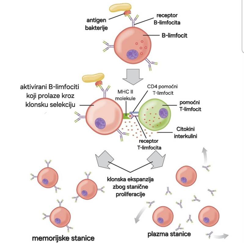
The image shows the mechanism of the immune response:
- a bacterial antigen is recognised by a B-lymphocyte receptor;
- the B-lymphocyte is activated;
- an MHC class II molecule presents the processed antigen and directs a specific immune response;
- the B-lymphocyte presents the bacterial antigen and releases cytokines;
- cytokines activate memory lymphocytes according to the antigen-receptor pattern;
- specific lymphocytes are cloned and attack the bacteria.
MHC class II
Macrophages and dendritic cells (DCs) are antigen-presenting cells that constitutively express MHC class II under steady-state conditions.
MHC class II molecules are expressed at high levels on activated T cells.
The MHC class II protein complex is encoded by the human leukocyte antigen (HLA) gene complex.
The most important function of human leukocyte antigen (HLA) molecules is to induce and regulate immune responses and select the T-lymphocyte repertoire.
In sarcoidosis and other autoimmune diseases, this function of HLA molecules is important in detecting an abnormal immune response.
Dino-Josip Ključarić